
What Can a Sinus CT Show That an Office Examination Cannot?
Persistent nasal congestion, facial pressure, postnasal drip, headaches, or recurring sinus infections can have several possible causes. An office examination is often the first step in determining what may be contributing to these symptoms. In some cases, however, an examination cannot provide a complete view of the deeper sinus cavities.
When additional information is needed, an ENT care provider may recommend a sinus CT scan. At SoCal Breathe Free, the clinical team helps patients in Burbank and the surrounding Los Angeles area understand which diagnostic tests may be appropriate based on their symptoms, examination findings, and medical history.
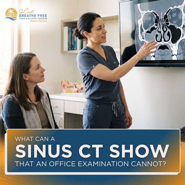
Direct Answer: What Does a Sinus CT Show?
A sinus CT produces detailed, cross-sectional images of the nasal passages, sinus cavities, drainage pathways, and surrounding bony structures. It may reveal inflammation, trapped fluid, narrowed drainage pathways, nasal polyps, or anatomical variations that cannot be fully evaluated during a standard office examination.
A CT scan complements an office examination; it does not replace one. The examination shows the appearance of directly visible nasal tissue, while CT imaging provides a broader view of structures located deeper inside the face.
What Is a Sinus CT Scan?
A computed tomography, or CT, scan is a diagnostic imaging examination that uses specialized X-ray equipment and computer processing to create cross-sectional images of the body.
A CT scan of the sinuses captures images of the air-filled sinus cavities located within the bones of the face. The images can be reviewed from multiple angles, allowing a qualified healthcare provider to evaluate the internal anatomy in greater detail than is possible with a conventional X-ray.
Sinus CT imaging may be used to:
- Evaluate persistent or recurrent sinus symptoms
- Look for fluid or thickened sinus tissue
- Assess whether sinus drainage pathways are narrowed or obstructed
- Identify certain structural variations
- Evaluate the extent of inflammatory sinus disease
- Help plan a procedure when clinically appropriate
- Investigate an abnormality that requires further evaluation
Routine sinus CT scans are commonly performed without contrast material. Contrast may be recommended when a provider needs additional information about certain complications, masses, blood vessels, or surrounding soft tissues.
What Happens During a Sinus CT?
The exact process depends on the equipment and facility. During a conventional CT scan, the patient generally lies on a table that moves through the scanner’s circular opening. The equipment takes a series of X-ray images from different angles, and a computer reconstructs them into detailed views of the sinuses.
Some ENT offices use specialized in-office CT equipment that allows the patient to sit or stand during the scan. The imaging process is generally brief and noninvasive, although the total appointment time may vary.
Patients should remain still while the images are captured. They should also tell their healthcare provider if they may be pregnant, have recently undergone other imaging examinations, or have concerns about radiation exposure.
What Can a Sinus CT Reveal That an Office Examination May Not?
An office examination and a sinus CT provide different types of information. Depending on the patient’s symptoms, CT imaging may reveal the following findings that cannot be completely assessed by looking inside the nose.
Deeper sinus cavities
An office examination can evaluate the entrance to the nose and directly visible portions of the nasal passages. Nasal endoscopy can extend that view and help an ENT provider examine drainage pathways and other areas inside the nose.
Even with an endoscope, however, the provider cannot see through bone or directly inspect every sinus cavity. A CT scan can show the:
- Maxillary sinuses in the cheek area
- Ethmoid sinuses between the eyes
- Frontal sinuses above the eyes
- Sphenoid sinuses deeper behind the nasal cavity
This broader view can help determine which sinuses appear affected and whether the findings are limited to one area or involve multiple sinus cavities.
Mucosal thickening and sinus opacification
The sinuses are normally filled with air. Inflammation can cause the tissue lining a sinus to thicken, while fluid or inflammatory material may partially or completely fill the cavity.
A sinus CT may show:
- Thickening of the sinus lining
- Fluid levels
- Partial or complete sinus opacification
- Patterns of inflammation affecting individual sinus cavities
These findings must be interpreted alongside the patient’s symptoms and examination. Imaging abnormalities do not always mean that a patient has an active bacterial infection, and mild changes may occasionally be found in people without significant sinus symptoms.
Narrowed or obstructed drainage pathways
Each sinus drains through a small opening. Swelling, structural narrowing, polyps, or other changes may interfere with drainage.
A CT scan can help map these pathways and show areas where normal ventilation or drainage may be restricted. This information may be useful when symptoms have persisted despite appropriate medical management or when a procedure is being considered.
Structural differences inside the nose and sinuses
Sinus anatomy differs from person to person. A CT scan may identify anatomical variations that are difficult or impossible to assess fully during an office examination.
These may include:
- A deviated nasal septum
- Enlarged turbinates
- Narrow sinus openings
- Variations in the small bony structures surrounding the sinuses
- Air cells that may affect a drainage pathway
The presence of an anatomical variation does not automatically mean it is causing symptoms or requires treatment. An ENT provider can determine whether a finding corresponds with the patient’s symptoms and clinical examination.
Nasal polyps and the extent of inflammatory disease
An office examination or nasal endoscopy may identify nasal polyps that extend into visible areas of the nasal passages. A CT scan may provide additional information about how extensively inflammatory tissue or polyps involve the deeper sinus cavities.
This can help a provider assess the overall pattern of disease and discuss treatment options appropriate to the individual patient.
Findings near the upper teeth
The roots of the upper teeth are located close to the maxillary sinuses. In some cases, dental disease or a prior dental procedure may be associated with inflammation in a nearby sinus.
A sinus CT may show findings that raise concern for a possible dental source, but it does not replace a dental examination or dedicated dental imaging. Additional evaluation by a dentist or oral healthcare professional may be recommended.
Uncommon abnormalities
CT imaging may occasionally reveal a mass, bone change, or another unexpected abnormality that requires additional assessment.
A sinus CT alone may not determine exactly what an abnormality represents. Depending on the finding, a provider may recommend nasal endoscopy, MRI, biopsy, or referral for further evaluation.
What Can an Office Examination Show?
A sinus CT provides important anatomical information, but it cannot replace the clinical information gathered during an office visit.
An office examination allows the provider to:
- Discuss the patient’s symptoms and medical history
- Examine the ears, nose, mouth, and throat
- Evaluate visible swelling, irritation, crusting, or bleeding
- Look for nasal drainage
- Assess airflow through each nostril
- Evaluate the effects of allergies or other inflammatory conditions
- Perform nasal endoscopy when appropriate
Nasal endoscopy provides a real-time view of accessible nasal tissue. It may identify inflammation, drainage, crusting, polyps, bleeding sites, or other visible abnormalities. Unlike a CT scan, it also allows the provider to assess how the tissue appears during the examination.
Because the two evaluations answer different questions, an ENT provider may use both when clinically appropriate.
Does Everyone With Sinus Symptoms Need a CT Scan?
No. A CT scan is not routinely necessary for every cold, episode of congestion, or uncomplicated case of acute sinusitis.
A provider may consider sinus CT imaging when:
- Symptoms have persisted despite appropriate treatment
- Sinus infections recur frequently
- Chronic rhinosinusitis is suspected
- The diagnosis remains unclear after an examination
- A complication is suspected
- Symptoms or examination findings are unusual
- Detailed anatomy is needed for procedure planning
CT imaging uses ionizing radiation. Although the radiation exposure from a medically appropriate scan is generally considered small, the examination should be ordered when its expected diagnostic benefit outweighs its potential risk. The provider may also consider previous imaging and whether another examination would answer the clinical question with less or no radiation.
When Should Persistent Sinus Symptoms Be Evaluated?
Consider scheduling an evaluation when nasal or sinus symptoms:
- Persist longer than expected
- Recur frequently
- Do not improve with appropriate treatment
- Interfere with breathing, sleep, work, or daily comfort
- Include ongoing facial pressure or reduced sense of smell
- Are consistently worse on one side
- Include repeated or unexplained bloody nasal discharge
Seek prompt medical attention for severe headache, swelling or redness around the eyes, vision changes, confusion, neck stiffness, significant facial swelling, difficulty breathing, or rapidly worsening symptoms.
Understanding the Role of Sinus CT Imaging
A sinus CT can provide information that an office examination cannot by showing the deeper sinus cavities, drainage pathways, surrounding bone, and overall pattern of sinus inflammation. However, the images are most useful when interpreted together with the patient’s symptoms, medical history, and physical examination.
SoCal Breathe Free provides ear, nose, and throat evaluations for patients in Burbank and the surrounding Los Angeles area. If persistent or recurring sinus symptoms are affecting your daily life, schedule an appointment with the Burbank team to discuss what may be contributing and whether additional testing may be appropriate.

It isn’t always obvious where to turn when you’re dealing with congestion, sinus pressure, sneezing, or a constantly stuffy nose. Many people assume…

Not every “cold” that lingers is actually a cold. If your congestion, sneezing, or itchy eyes seem to return around the same time each year, there’s…